The uncertainty you feel while waiting for biopsy test results can cause a lot of stress and anxiety.
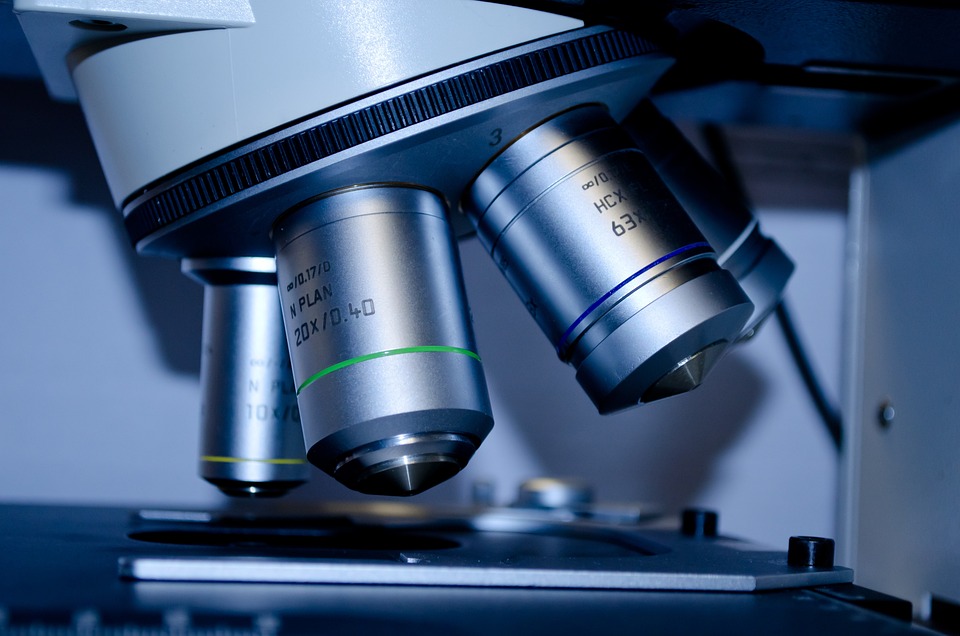
To determine if you have cancer or another condition, the doctor removes a sample of abnormal tissue for a biopsy. A pathologist looks at the tissue under a microscope and runs other tests on the cells. The pathologist describes the findings on the tissue’s health in a pathology report.
While this may sound like a simple process, it isn’t!
The slicing and staining of a sample and inspecting it microscopically is a very resource-intensive procedure that requires the expertise of trained pathologists.
Waiting for a pathologist’s report may soon be a thing of the past!
A team of researchers developed a quick and simple method of accurately analyzing solid cancer tumors in about 30 minutes without consulting a pathologist.
Scientists from the Max Planck Institute for the Science of Light (MPL) and FAU have created a fast and precise method for analyzing cells in tissue samples using artificial intelligence.
The new method uses a “tissue grinder” to tear apart biopsy samples down to single cells, a process that takes less than five minutes.
These single cells are then analyzed using the real-time deformability cytometry (RT-DC) method developed in the Guck laboratory. The AI-enabled model evaluates the data obtained by RT-FDC and quickly assesses whether a biopsy sample contains tumor cells.
RT-FDC can analyze up to 1,000 cells per second which is 36,000 times faster than traditional methods of analyzing cell deformability.